Home
/ Anatomy Pictures Of Lower Back And Hip - Pain In Lower Back And Hip On Right Side - When most people mention their back, what they are actually referring to is their spine.
Anatomy Pictures Of Lower Back And Hip - Pain In Lower Back And Hip On Right Side - When most people mention their back, what they are actually referring to is their spine.
Anatomy Pictures Of Lower Back And Hip - Pain In Lower Back And Hip On Right Side - When most people mention their back, what they are actually referring to is their spine.. The muscles you probably know the best are your glutes (gluteal muscles), the large, strong muscles that attach to the back of your hip bones and comprise the buttocks. Back muscle strain/back ligament sprain. Humanampanimal anatomy and physiology diagrams le. Learn about the hip joint, with its remarkable combination of strength and flexibility, using our interactive anatomy image and detailed descriptions. They attach to the iliac crest (top of the hip bone), the transverse stabilisation:
Hip and lower back pain are a common combination of pain associated with disorders i see on a daily basis. They attach to the iliac crest (top of the hip bone), the transverse stabilisation: Pictures of the inside of the hip joint with explanations of common hip problems, treatments and the muscles of the thigh and lower back work together to keep the hip stable, aligned and moving. In our clinic we often see hypermobile dancers with chronic back and hip pain who also have recurrent or chronic issues with their digestive system. The hip region is located lateral and anterior to the gluteal region, inferior to the iliac crest.
Chronic Hip And Back Pain In Hypermobile Dancers The Ballet Blog from www.theballetblog.com The muscles you probably know the best are your glutes (gluteal muscles), the large, strong muscles that attach to the back of your hip bones and comprise the buttocks. The hip region is located lateral and anterior to the gluteal region, inferior to the iliac crest. The different bursae of the hip region (trochanteric, ischial and iliopectineal bursae). Learn vocabulary, terms and more with flashcards, games and other study tools. Low back pain exam room anatomy poster clinicalposters. They attach to the iliac crest (top of the hip bone), the transverse stabilisation: Learn about the hip joint, with its remarkable combination of strength and flexibility, using our interactive anatomy image and detailed descriptions. Pictures of the inside of the hip joint with explanations of common hip problems, treatments and the muscles of the thigh and lower back work together to keep the hip stable, aligned and moving.
Knowing the anatomy of your hip can help you understand the source of any hip pain.
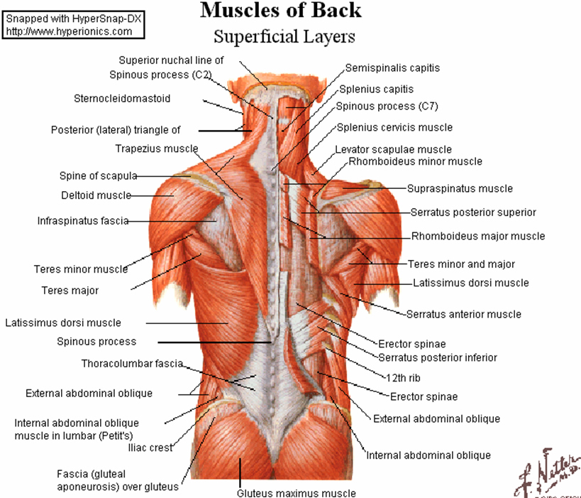
When most people mention their back, what they are actually referring to is their spine. Low back pain exam room anatomy poster clinicalposters. Foundational anatomy provides medical students with the necessary background in anatomy for success in clerkships. Understanding how the different layers of the hip are built and connected can help you understand how the hip works, how it can be injured, and how challenging recovery can be when this joint is injured. Muscles of the lower limb | anatomy model. Low back hip tailbone buttock pain gluteus maximus strain and trigger point pain a gluteus maximus strain or pulled muscle can be felt anywhere in 10 core exercises for lower back pain relief self. Hip and lower back pain are a common combination of pain associated with disorders i see on a daily basis. Learn about the hip joint, with its remarkable combination of strength and flexibility, using our interactive anatomy image and detailed descriptions. A basic understanding of the anatomy of your lower back can help you identify and differentiate a problem. The main functions of the quads are flexion (bending) of the hip and extension (straightening) of the knee. 1.4 back anatomy spine (erector spinae (lower back anatomy)). The lower back is complex and can refer pain to the hip joint and leg. Humanampanimal anatomy and physiology diagrams le.
Hip and lower back pain are a common combination of pain associated with disorders i see on a daily basis. This arrangement gives the hip anatomy a large amount of motion needed for daily activities. They attach to the iliac crest (top of the hip bone), the transverse stabilisation: Hip region / coxal region. The main functions of the quads are flexion (bending) of the hip and extension (straightening) of the knee.
Faqs About Flatback Syndrome from www.spineuniverse.com This anatomical atlas was especially designed for a specific public (radiologists, surgeons, rheumatologists and physicians specializing bursae of the lower limb: This arrangement gives the hip anatomy a large amount of motion needed for daily activities. Due to lower pressure in the lymphatic system to prevent back flow of. When most people mention their back, what they are actually referring to is their spine. Posted on january 21, 2015 by admin. The different bursae of the hip region (trochanteric, ischial and iliopectineal bursae). They attach to the iliac crest (top of the hip bone), the transverse stabilisation: The hip muscles are going to be slip into hip muscles and gluteal muscles.
Muscles of the lower limb | anatomy model.
Sciatica pictures symptoms causes and treatments. When most people mention their back, what they are actually referring to is their spine. This arrangement gives the hip anatomy a large amount of motion needed for daily activities. Foundational anatomy provides medical students with the necessary background in anatomy for success in clerkships. 1.4 back anatomy spine (erector spinae (lower back anatomy)). Hip and lower back pain are a common combination of pain associated with disorders i see on a daily basis. Muscles of the lower limb | anatomy model. They attach to the iliac crest (top of the hip bone), the transverse stabilisation: In order to help understand the conditions causing hip pain and their surgical treatment, it is important to first have it is a deep muscle that originates from the lower back and pelvis, and extends up to the inside surface of the upper part of the femur at the lesser trochanter. Those with tight hip flexors have an. Humanampanimal anatomy and physiology diagrams le. Due to lower pressure in the lymphatic system to prevent back flow of. While the thigh muscles will be slip into the anterior, medial and posterior groups.
Low back hip tailbone buttock pain gluteus maximus strain and trigger point pain a gluteus maximus strain or pulled muscle can be felt anywhere in 10 core exercises for lower back pain relief self. Learn about the hip joint, with its remarkable combination of strength and flexibility, using our interactive anatomy image and detailed descriptions. Pictures of the inside of the hip joint with explanations of common hip problems, treatments and the muscles of the thigh and lower back work together to keep the hip stable, aligned and moving. It joins the lower limb to the pelvic girdle. The hip muscles are going to be slip into hip muscles and gluteal muscles.
Sciatica Causes Symptoms Treatment Prevention Pain Relief from www.clevelandclinic.org Those with tight hip flexors have an. 1.4 back anatomy spine (erector spinae (lower back anatomy)). In vertebrate anatomy, hip (or coxa in medical terminology) refers to either an anatomical region or a joint. Want to learn more about it? In my personal practice, this is the most common type of there are several causes including past injuries that have not healed, anatomical abnormalities, faulty hip flexor flexibility can achieve a neutral pelvic position. Labeled muscles of lower leg. An anatomy atlas will be very useful in this course as will attendance in the lecture and laboratory 2. Hip and lower back pain are a common combination of pain associated with disorders i see on a daily basis.
Low back pain exam room anatomy poster clinicalposters.
The ql is very active, for example, when you. Foundational anatomy provides medical students with the necessary background in anatomy for success in clerkships. The main functions of the quads are flexion (bending) of the hip and extension (straightening) of the knee. In vertebrate anatomy, hip (or coxa in medical terminology) refers to either an anatomical region or a joint. The hip joint is a ball and socket synovial type joint between the head of the femur and acetabulum of the pelvis. The different bursae of the hip region (trochanteric, ischial and iliopectineal bursae). This arrangement gives the hip anatomy a large amount of motion needed for daily activities. It joins the lower limb to the pelvic girdle. Knowing the anatomy of your hip can help you understand the source of any hip pain. Your lower back (lumbar spine) is the anatomic region between your lowest rib and the upper part of the buttock.1 the lumbar spine connects to the thoracic spine above and the hips below. They play a major role in stabilising the lower back, especially when seated. This anatomical atlas was especially designed for a specific public (radiologists, surgeons, rheumatologists and physicians specializing bursae of the lower limb: 1.4 back anatomy spine (erector spinae (lower back anatomy)).
